These medical illustrations were created for a patient education flip book about gynecologic health and gynecologic cancers.
#medicalillustration #illustrationsmedical #medart #medicalartwork #sciart #biologicalart #medicalcommunication #anatomyart #anatomicalart #artandanatomy #anatomydrawing #biologicalillustration #anatomyillustration #drawinganatomy #scienceart #scienceartist #scienceandart #medicalartist #medicaleducation #medicalstudent #meded #scicomm #scicommunity #healthcareart #patienteducation #gynecologicalhealth #Cervicalcancer #gynecology #womenshealthmatters #courtneyardenstudio
This medical illustration is about cardiac electricity axis deviation, created for medical education. The heart has an electrical conduction system that allows the heart to keep beating. The cardiac axis describes the overall direction as the cardiac electricity spreads through the system. A healthy person would be between -30 and 90 degrees. Several conditions can cause the axis to deviate to the right (RAD) or left (LAD), including an enlarged heart ventricle, myocardial infarction, congenital heart disease, and hyperkalemia.
#medicalillustration #illustrationsmedical #medart #medicalartwork #sciart #biologicalart #medicalcommunication #anatomyart #anatomicalart #artandanatomy #anatomydrawing #biologicalillustration #anatomyillustration #drawinganatomy #scienceart #scienceartist #scienceandart #medicalartist #medicaleducation #medicalstudent #meded #scicomm #scicommunity #healthcareart #patienteducation #cardiology #heartdisease #cardiacconduction #myocardialinfarction #courtneyardenstudio
During diastole, the atrioventricular valves open to allow the ventricles to fill with blood. They close during systole when the ventricles contract to direct blood through the semilunar valves to the body. When the valve opening narrows, it is called stenosis, a valve disease restricting blood flow. Regurgitation is when the valve doesn't close completely, and the blood can leak backward.
#medicalillustration #illustrationsmedical #medart #medicalartwork #sciart #biologicalart #medicalcommunication #anatomyart #anatomicalart #artandanatomy #anatomydrawing #biologicalillustration #anatomyillustration #drawinganatomy #scienceart #scienceartist #scienceandart #medicalartist #medicaleducation #medicalstudent #meded #scicomm #scicommunity #healthcareart #patienteducation #heartvalvedisease #hearthealth #cardiologist #heartvalve #courtneyardenstudio
This medical illustration shows the different patterns of myocardial infarctions. A myocardial infarction is a heart attack that happens when blood flow to the heart is blocked, which causes parts of the muscle of the heart to die.
#medicalillustration #illustrationsmedical #medart #medicalartwork #sciart #biologicalart #medicalcommunication #anatomyart #anatomicalart #artandanatomy #anatomydrawing #biologicalillustration #anatomyillustration #drawinganatomy #scienceart #scienceartist #scienceandart #medicalartist #medicaleducation #medicalstudent #meded #scicomm #scicommunity #healthcareart #patienteducation #hearthealthawareness #heartdiseaseawareness #cardiology #myocardialinfarction #courtneyardenstudio
This medical illustration is about the semilunar valves in the heart, comparing proper function, stenosis, and regurgitation. The semilunar valves control blood flow from the ventricles to the arteries, preventing blood from flowing backward. Stenosis is when the valves narrow and can't fully open. Regurgitation is when the valves don't close completely, and blood flows backward.
#medicalillustration #illustrationsmedical #medart #medicalartwork #sciart #biologicalart #medicalcommunication #anatomyart #anatomicalart #artandanatomy #anatomydrawing #biologicalillustration #anatomyillustration #drawinganatomy #scienceart #scienceartist #scienceandart #medicalartist #medicaleducation #medicalstudent #meded #scicomm #scicommunity #healthcareart #patienteducation #heartdiseaseawareness #hearthealth #heartvalvedisease #valveregurgitation #courtneyardenstudio
This medical illustration is about mitral regurgitation, which is a heart condition that happens when the mitral valve doesn't close properly, causing blood to flow backward from the left ventricle to the left atrium. Less blood then gets pumped to the rest of the body.
#medicalillustration #illustrationsmedical #medart #medicalartwork #sciart #biologicalart #medicalcommunication #anatomyart #anatomicalart #artandanatomy #anatomydrawing #biologicalillustration #anatomyillustration #drawinganatomy #scienceart #scienceartist #scienceandart #medicalartist #medicaleducation #medicalstudent #meded #scicomm #scicommunity #healthcareart #patienteducation #mitralregurgitation #heartdiseases #mitralvalvedisease #hearthealth #courtneyardenstudio
This medical illustration is about transmural infarction vs nontransmural infarction. Transmural infarctions involve the entire thickness of the heart muscle wall from the endocardium to the epicardium, while nontransmural infarctions do not.
#medicalillustration #illustrationsmedical #medart #medicalartwork #sciart #biologicalart #medicalcommunication #anatomyart #anatomicalart #artandanatomy #anatomydrawing #biologicalillustration #anatomyillustration #drawinganatomy #scienceart #scienceartist #scienceandart #medicalartist #medicaleducation #medicalstudent #meded #scicomm #scicommunity #healthcareart #patienteducation #hearthealth #myocardialinfarction #cardiology #heartanatomy ##courtneyardenstudio
This medical illustration shows the flow of cerebrospinal fluid (CSF) through the brain ventricles. It circulates around the brain and spinal cord in the subarachnoid space, and it is removed by the arachnoid granulations in the superior sagittal sinus, where it enters the bloodstream.
#medicalillustration #illustrationsmedical #medart #medicalartwork #sciart #biologicalart #medicalcommunication #anatomyart #anatomicalart #artandanatomy #anatomydrawing #biologicalillustration #anatomyillustration #drawinganatomy #scienceart #scienceartist #scienceandart #medicalartist #medicaleducation #medicalstudent #meded #scicomm #scicommunity #healthcareart #patienteducation #Neuroanatomy #Brainillustration, #cerebralspinalfluid, #Brainhealth, #courtneyardenstudio
This medical illustration is of the choroid plexus, an area in the brain's ventricles comprised of networks of fenestrated capillaries and cells. It forms a big part of the blood-brain barrier and produces cerebral spinal fluid via the ependymal cells surrounding the capillaries and connective tissue.
#medicalillustration #illustrationsmedical #medart #medicalartwork #sciart #biologicalart #medicalcommunication #anatomyart #anatomicalart #artandanatomy #anatomydrawing #biologicalillustration #anatomyillustration #drawinganatomy #scienceart #scienceartist #scienceandart #medicalartist #medicaleducation #medicalstudent #meded #scicomm #scicommunity #healthcareart #patienteducation #choroidplexuscarcinoma #braincells #astrocytes #brainart #courtneyardenstudio
This medical illustration shows different types of endocarditis in the heart. Endocarditis is inflammation of the endocardium, the lining of the heart's valves and chambers. It occurs mostly from bacteria that travel through the bloodstream from somewhere else in the body and attach to the endocardium. Nonbacterial endocarditis is associated with cancer or autoimmune disorders. Libman-Sacks Endocarditis is a type of nonbacterial thrombotic endocarditis associated with systemic lupus erythematosus.
#medicalillustration #illustrationsmedical #medart #medicalartwork #sciart #biologicalart #medicalcommunication #anatomyart #anatomicalart #artandanatomy #anatomydrawing #biologicalillustration #anatomyillustration #drawinganatomy #scienceart #scienceartist #scienceandart #medicalartist #medicaleducation #medicalstudent #meded #scicomm #scicommunity #healthcareart #patienteducation #Endocarditis
#endocarditissurvivor #heartdiseases #heartanatomy #courtneyardenstudio
These illustrations were created for a defense case. The first illustration shows chronic changes in the lumbar spine compared to what would be expected from an acute traumatic injury. The second illustration shows the cervical spine with degenerative changes compared to the cervical spine with acute traumatic injuries.
#medicalillustration #illustrationsmedical #medart #medicalartwork #sciart #biologicalart #medicalcommunication #anatomyart #anatomicalart #artandanatomy #anatomydrawing #biologicalillustration #anatomyillustration #drawinganatomy #scienceart #scienceartist #scienceandart #medicalartist #medicaleducation #medicalstudent #meded #scicomm #scicommunity #healthcareart #patienteducation #medicallegal #courtroomillustration #spinalhealth #backinjuries #courtneyardenstudio
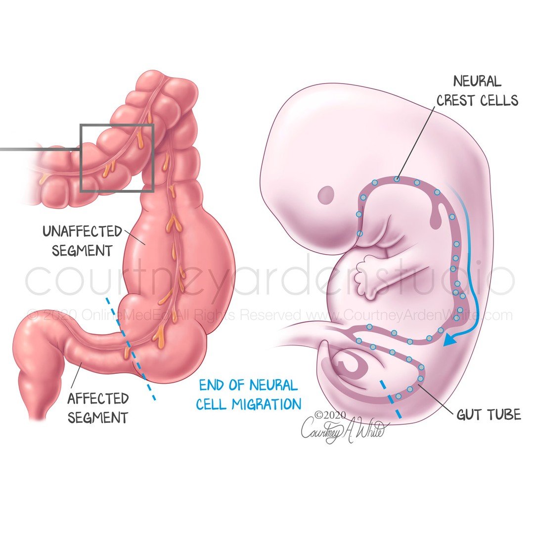
This illustration is about Hirschsprung disease (HSCR,aganglionic megacolon), a congenital disease that affects the colon. During development, the neural crest cells cease migrating, preventing nerves from forming in part of the colon (large intestine). This causes the healthy segment of colon to swell with stool that can't pass through the segment of colon without nerves.
#medicalillustration #illustrationsmedical #medart #medicalartwork #sciart #biologicalart #medicalcommunication #anatomyart #anatomicalart #artandanatomy #anatomydrawing #biologicalillustration #anatomyillustration #drawinganatomy #scienceart #scienceartist #scienceandart #medicalartist #medicaleducation #medicalstudent #meded #scicomm #scicommunity #healthcareart #patienteducation #megacolon #Colonhealth #gastrointestinalproblems #hirschsprungsdisease #courtneyardenstudio
This medical illustration is about cervical effacement and dilation during pregnancy and labor, which allows the baby to descend through the birth canal. Effacement or ripening is when the cervix softens, thins, and shortens. Dilation is the opening of the cervix from 0cm to 10cm. When the cervix is 10cm dilated and 100%, it is ready for delivery.
#medicalillustration #illustrationsmedical #medart #medicalartwork #sciart #biologicalart #medicalcommunication #anatomyart #anatomicalart #artandanatomy #anatomydrawing #biologicalillustration #anatomyillustration #drawinganatomy #scienceart #scienceartist #scienceandart #medicalartist #medicaleducation #medicalstudent #meded #scicomm #scicommunity #healthcareart #patienteducation #midwiferystudent #obstetrics #birthcenter #laboranddelivery #courtneyardenstudio
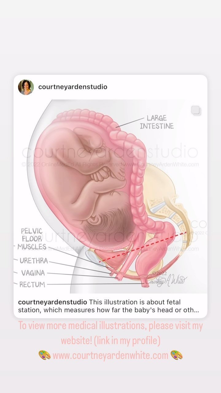
This illustration is about fetal station, which measures how far the baby's head or other presenting part of the baby's body has descended into the mother's pelvis during labor. It begins with station -5. Zero is at the level of the ischial spine of the pelvis, and +5 is the station at birth.
If you want to view more medical illustrations about pregnancy, obstetrics or any other medical subject, please visit my website! (link in my profile)
🎨 www.courtneyardenwhite.com 🎨
#medicalillustration #illustrationsmedical #medart #medicalartwork #sciart #biologicalart #medicalcommunication #anatomyart #anatomicalart #artandanatomy #anatomydrawing #biologicalillustration #anatomyillustration #drawinganatomy #scienceart #scienceartist #scienceandart #medicalartist #medicaleducation #medicalstudent #meded #scicomm #scicommunity #healthcareart #patienteducation #fetalstation #obstetricsandgynecology #midwiferystudent #midwiferymodelofcare #courtneyardenstudio
This illustration is about gestational trophoblastic disease (GTD) and neoplasm. GTD is rare and occurs when abnormal trophoblast cells grow inside the uterus after conception. Trophoblast cells surround the fertilized egg and eventually make up a large part of the placenta. However, in rare cases, if there is a problem with these cells, a tumor can develop instead of a fetus. Most GTD is not cancerous, but some can become cancer and spread to other parts of the body. GTD is a term used to describe different types of disease, including a partial mole, complete mole, invasive mole, choriocarcinoma, and very rare, placental-site trophoblastic tumor or epithelioid trophoblastic tumor.
If you're interested in viewing more medical illustrations about reproduction and pregnancy or any other medical subject, please visit my website. (link in my profile)
🎨 www.courtneyardenwhite.com 🎨
#medicalillustration #illustrationsmedical #medart #medicalartwork #sciart #biologicalart #medicalcommunication #anatomyart #anatomicalart #artandanatomy #anatomydrawing #biologicalillustration #anatomyillustration #drawinganatomy #scienceart #scienceartist #scienceandart #medicalartist #medicaleducation #medicalstudent #meded #scicomm #scicommunity #healthcareart #patienteducation #womenshealthmatters #abnormalpregnancy #pregnancydiseases #reproductivehealthcare #courtneyardenstudio
If you're interested in viewing more medical illustrations about the lungs or any other medical subject, please visit my website! (link in my profile)
🎨 www.courtneyardenwhite.com 🎨